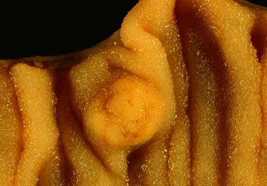
ଫାଇଲ:Multiple Carcinoid Tumors of the Small Bowel 2.jpg
Multiple_Carcinoid_Tumors_of_the_Small_Bowel_2.jpg (୫୫୦ × ୩୮୪ ପିକସେଲ, ଫାଇଲ ଆକାର: ୩୨ KB, ଏମ.ଆଇ.ଏମ.ଇର ପ୍ରକାର: image/jpeg)
ଫାଇଲ ଇତିହାସ
ଏହା ଫାଇଲଟି ସେତେବେଳେ ଯେମିତି ଦିଶୁଥିଲା ତାହା ଦେଖିବା ପାଇଁ ତାରିଖ/ବେଳା ଉପରେ କ୍ଲିକ କରନ୍ତୁ
| ତାରିଖ/ବେଳ | ନଖ ଦେଖଣା | ଆକାର | ବ୍ୟବହାରକାରୀ | ମତାମତ | |
|---|---|---|---|---|---|
| ଏବେକାର | ୦୪:୩୬, ୫ ଜୁନ ୨୦୦୬ |  | ୫୫୦ × ୩୮୪ (୩୨ KB) | Patho | {{Information| |Description=The photo shows the same case presented in [http://commons.wikimedia.org/wiki/Image:Multiple_Carcinoid_Tumors_of_the_Small_Bowel_1.jpg Image:Multiple_Carcinoid_Tumors_of_the_Small_Bowel_1.jpg]. The medium close-up photo above |
ଫାଇଲ ବ୍ୟବହାର
ଏହି ସବୁପୃଷ୍ଠା ଏହି ଫାଇଲଟିକୁ ଯୋଡ଼ିଥାନ୍ତି:
ଜଗତ ଫାଇଲ ବ୍ୟବହାର
ତଳଲିଖିତ ଉଇକିସବୁ ଏହି ଫାଇଲଟିକୁ ବ୍ୟବହାର କରିଥାନ୍ତି:
- ar.wikipedia.orgରେ ବ୍ୟବହାର
- az.wikipedia.orgରେ ବ୍ୟବହାର
- de.wikipedia.orgରେ ବ୍ୟବହାର
- de.wikibooks.orgରେ ବ୍ୟବହାର
- en.wikipedia.orgରେ ବ୍ୟବହାର
- es.wikipedia.orgରେ ବ୍ୟବହାର
- fr.wikipedia.orgରେ ବ୍ୟବହାର
- he.wikipedia.orgରେ ବ୍ୟବହାର
- hy.wikipedia.orgରେ ବ୍ୟବହାର
- it.wikipedia.orgରେ ବ୍ୟବହାର
- ko.wikipedia.orgରେ ବ୍ୟବହାର
- pl.wikipedia.orgରେ ବ୍ୟବହାର
- pt.wikipedia.orgରେ ବ୍ୟବହାର
- sr.wikipedia.orgରେ ବ୍ୟବହାର
- sv.wikipedia.orgରେ ବ୍ୟବହାର
- ta.wikipedia.orgରେ ବ୍ୟବହାର
- uk.wikipedia.orgରେ ବ୍ୟବହାର
- www.wikidata.orgରେ ବ୍ୟବହାର
- zh.wikipedia.orgରେ ବ୍ୟବହାର
