ଫାଇଲ:Minimal Change Disease Pathology Diagram.svg

Size of this PNG preview of this SVG file: ୬୨୮ × ୬୦୦ ପିକ୍ସେଲ. ବାକି ରେଜୋଲୁସନ: ୨୫୧ × ୨୪୦ ପିକ୍ସେଲ | ୫୦୩ × ୪୮୦ ପିକ୍ସେଲ | ୮୦୪ × ୭୬୮ ପିକ୍ସେଲ | ୧,୦୭୨ × ୧,୦୨୪ ପିକ୍ସେଲ | ୨,୧୪୪ × ୨,୦୪୮ ପିକ୍ସେଲ | ୭୧୫ × ୬୮୩ ପିକ୍ସେଲ.
ମୂଳ ଫାଇଲ (SVG ଫାଇଲ, ସାଧାରଣ ମାପ ୭୧୫ × ୬୮୩ ପିକ୍ସେଲ, ଫାଇଲ ଆକାର: ୧୮୬ KB)
This is a file from the Wikimedia Commons. Information from its description page there is shown below. |
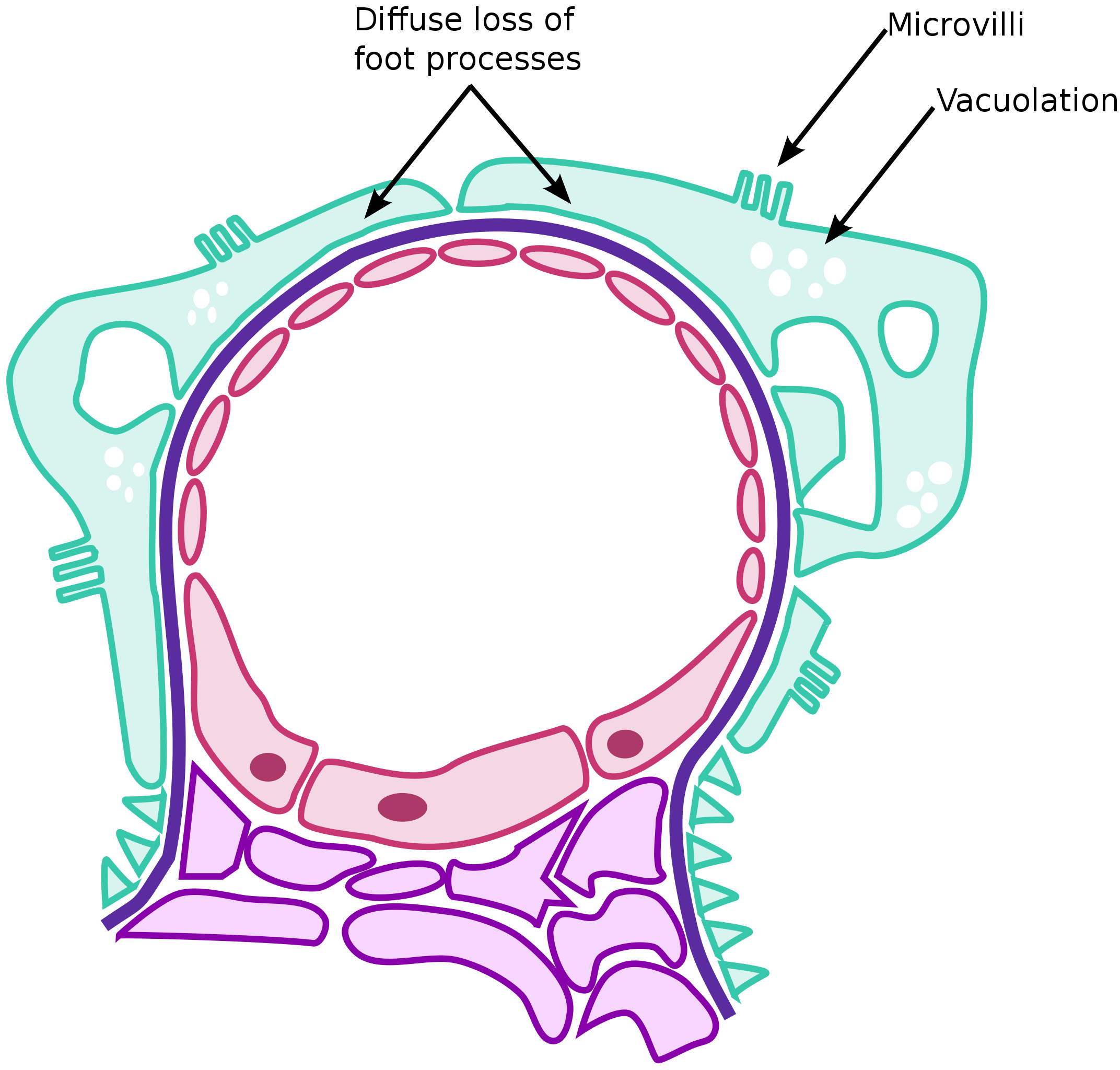
| ବିବରଣୀ | A schematic of the changes seen under the electron microscope of minimal change disease. |
| ତାରିଖ | (UTC) |
| ମୂଳାଧାର | |
| ଲେଖକ |
|
| This is a retouched picture, which means that it has been digitally altered from its original version. Modifications: pathology of minimal change disease. The original can be viewed here: Renal corpuscle.svg:
|
ମୁଁ, ଏହି କାମର ସତ୍ଵାଧିକାରୀ, ଏଠାରେ ତଳଲିଖିତ ଲାଇସେନ୍ସ ଅଧୀନରେ ଏହାକୁ ପ୍ରକାଶ କଲି:
ଏହି ଫାଇଲଟି କ୍ରିଏଟିଭ କମନ୍ସ ଅଧୀନରେ ଆଟ୍ରିବୁସନ ସେଆର-ଏଲାଇକ ୩.୦ ଅନପୋର୍ଟେଡ଼ ଲାଇସେନ୍ସରେ ପଞ୍ଜିକରଣ କରାଯାଇଅଛି ।
- ଆପଣ ଆରାମରେ:
- ବାଣ୍ଟିପାରିବେ – କାମଟିକୁ ନକଲ କରିପାରିବେ, ବାଣ୍ଟିପାରିବେ ଓ ପ୍ରସାରଣ କରିପାରିବେ
- ମିଶାଇପାରିବେ – କାମଟି ଅଭିଯୋଜନ କରିପାରିବେ
- ତଳଲିଖିତ ସର୍ତ୍ତାବଳୀ ଅଧୀନରେ:
- ଶ୍ରେୟ – ଆପଣ ମନେ କରି ଏହି କାମର ଆବଶ୍ୟକୀୟ ଶ୍ରେୟ ମୂଳ ଗଢ଼ାଳି ବା ସ୍ୱତ୍ୱାଧୀକାରୀଙ୍କୁ ଦେବେ ଏବଂ ଦେଲାବେଳେ ଲାଇସେନ୍ସର ଲିଙ୍କ ଦେଇ କି କି ବଦଳ କଲେ ଉଲ୍ଲେଖ କରିବେ । ଏହା ଉପଯୁକ୍ତ ଢଙ୍ଗରେ କରିବେ କିନ୍ତୁ ଲାଇସେନ୍ସ ଦେଉଥିବା ବ୍ୟକ୍ତି ଆପଣଙ୍କ ପ୍ରଚାର କଲା ଭଳି କିଛି ଲେଖିବେ ନାହିଁ ।
- ସେଆର ଏଲାଇକ – ଯଦି ଆପଣ ଏହି କାମଟିକୁ ବଦଳାଇବେ, ରୂପାନ୍ତରଣ କରିବେ ବା ଏହାକୁ ନେଇ କିଛି ଗଢ଼ିବେ ତେବେ ଆପଣ ଏହାକୁ ଏକା ବା ଅଲଗା ଲାଇସେନ୍ସ ଭିତରେ ରଖିପାରିବେ ।
This license is only chosen due to the derivative nature of my work. All my work is in the public domain as I am both an intellectual property abolitionist and paid by the US federal government to be a medical student.
ମୂଲ ଅପଲୋଡ଼ ଫାଇଲ
This image is a derivative work of the following images:
- File:Renal_corpuscle.svg licensed with Cc-by-sa-3.0
- 2009-01-18T21:28:41Z M.Komorniczak 1030x760 (547361 Bytes) {{Information |Description= |Source= |Date= |Author= |Permission= |other_versions= }}
- 2009-01-18T19:14:24Z M.Komorniczak 1030x760 (545858 Bytes) == Opis == {{Information |Description={{pl|1=Schemat budowy [[:pl:ciałko nerkowe|ciałka nerkowego]] A - Ciałko nerkowe B - Kanalik proksymalny (kanalik główny) C - Kanalik dystalny (wstawka) D - Aparat przykłę
- 2009-01-16T18:34:11Z Eusebius 1030x760 (369837 Bytes) watermark removed [[Commons:Watermarks]]
- 2009-01-04T00:51:05Z M.Komorniczak 1030x760 (399333 Bytes) == Opis|Information == {{Information |Description={{pl|1=Schemat budowy [[:pl:ciałko nerkowe|ciałka nerkowego]] A - Ciałko nerkowe B - Kanalik proksymalny (kanalik główny) C - Kanalik dystalny (wstawka) D - Aparat
- 2008-11-10T23:05:18Z M.Komorniczak 665x515 (421026 Bytes) {{Information |Description={{pl|1=Schemat budowy [[:pl:ciałko nerkowe|ciałka nerkowego]] A - Ciałko nerkowe B - Kanalik proksymalny (kanalik główny) C - Kanalik dystalny (wstawka) 1. Błona podstawna 2. Część trzewna
Uploaded with derivativeFX
ଫାଇଲ ଇତିହାସ
ଏହା ଫାଇଲଟି ସେତେବେଳେ ଯେମିତି ଦିଶୁଥିଲା ତାହା ଦେଖିବା ପାଇଁ ତାରିଖ/ବେଳା ଉପରେ କ୍ଲିକ କରନ୍ତୁ
| ତାରିଖ/ବେଳ | ନଖ ଦେଖଣା | ଆକାର | ବ୍ୟବହାରକାରୀ | ମତାମତ | |
|---|---|---|---|---|---|
| ଏବେକାର | ୨୩:୪୩, ୨୧ ଫେବୃଆରୀ ୨୦୧୦ | | ୭୧୫ × ୬୮୩ (୧୮୬ KB) | Huckfinne | {{Information |Description=A schematic of the changes seen under the electron microscope of minimal change disease. |Source=*File:Renal_corpuscle.svg |Date=2010-02-21 18:00 (UTC) |Author=*File:Renal_corpuscle.svg: [[User:M.Komorniczak|<font co |
ଫାଇଲ ବ୍ୟବହାର
ଏହି ସବୁପୃଷ୍ଠା ଏହି ଫାଇଲଟିକୁ ଯୋଡ଼ିଥାନ୍ତି:
ଜଗତ ଫାଇଲ ବ୍ୟବହାର
ତଳଲିଖିତ ଉଇକିସବୁ ଏହି ଫାଇଲଟିକୁ ବ୍ୟବହାର କରିଥାନ୍ତି:
- de.wikipedia.orgରେ ବ୍ୟବହାର
- en.wikipedia.orgରେ ବ୍ୟବହାର
- es.wikipedia.orgରେ ବ୍ୟବହାର
- hr.wikipedia.orgରେ ବ୍ୟବହାର
- id.wikipedia.orgରେ ବ୍ୟବହାର
- it.wikipedia.orgରେ ବ୍ୟବହାର
- sv.wikipedia.orgରେ ବ୍ୟବହାର
- zh.wikipedia.orgରେ ବ୍ୟବହାର